Pelvic Anatomy Xray : Female pelvis bones and joints, X-ray - Stock Image C033 ... : It first appears too complicated to read the.
Pelvic Anatomy Xray : Female pelvis bones and joints, X-ray - Stock Image C033 ... : It first appears too complicated to read the.. Pelvis anatomy the pelvis is either the lower part of the trunk of the human body between the abdomen and the thighs. Drawn over a fractured hip fractures. Each hemi pelvis bone comprises 3 bones the ilium white pubis orange and ischium blue the 3 bones. Functional anatomy of the male. This video covers the following:
We are pleased to provide you with the picture named pelvis x ray anatomy. Pelvic anatomy mri variant anatomy pelvic viscera. Documents similar to systematic review of pelvical xray. Drawn over a fractured hip fractures. It first appears too complicated to read the.
Female Pelvis X-Ray Royalty Free Stock Photos - Image: 7064298 from thumbs.dreamstime.com Systematically examine all bony structures of the pelvis and femurs for symmetry, cortical breaks and joint spaces (sacroiliac, hip and. This mri male pelvis axial cross sectional anatomy tool is absolutely free to use. Drawn over a fractured hip fractures. ●to describe the approach for safe laparoscopic dissection. Functional anatomy of the male. The true pelvis is divided into three regions known as the pelvic brim, the cavity and the outlet. Pelvis anatomy the pelvis is either the lower part of the trunk of the human body between the abdomen and the thighs. The measurements of each of these regions is important as the fetal head has to negotiate its way through.
Branches of the internal iliac artery.
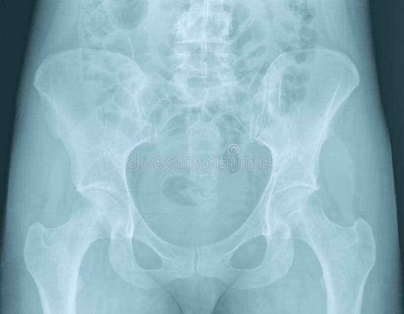
Documents similar to systematic review of pelvical xray. Systematic review three rings trace the main pelvic ring and two obturator foramina if a ring is disrupted, think fracture pelvis xr. This mri male pelvis axial cross sectional anatomy tool is absolutely free to use. Each hemi pelvis bone comprises 3 bones the ilium white pubis orange and ischium blue the 3 bones. Siu/icud consultation on urethral strictures: Drawn over a fractured hip fractures. Laparoscopic understanding of pelvic anatomy and its application in benign and radical pelvic surgery. There are many organs that sit in the pelvis, including much of the urinary system, and lots of the male or female reproductive systems. Surgical pelvic anatomy in gynecologic oncology. Learn vocabulary, terms and more with flashcards only rub 220.84/month. An x ray of the pelvis focuses specifically on the area between your hips that holds many of your reproductive and digestive organs. The true pelvis is divided into three regions known as the pelvic brim, the cavity and the outlet. The measurements of each of these regions is important as the fetal head has to negotiate its way through.
Siu/icud consultation on urethral strictures: Branches of the internal iliac artery. Identify the most important anatomical‐surgical landmarks on a pelvic model. Related online courses on physioplus. Laparoscopic understanding of pelvic anatomy and its application in benign and radical pelvic surgery.
Normal pelvic radiograph - female | Image | Radiopaedia.org from images.radiopaedia.org Branches of the internal iliac artery. Drawn over a fractured hip fractures. Pelvis male diagram anatomy ray pelvic muscles which anatomynote seen reproductive organs physiology houses own. Related online courses on physioplus. Learn vocabulary, terms and more with flashcards only rub 220.84/month. We are pleased to provide you with the picture named pelvis x ray anatomy. Unfortunately, the indirect view onto the anatomy in addition to projective simplication substantially. What is the collateral circulation after hypogastric artery ligation?
Drawn over a fractured hip fractures.
It first appears too complicated to read the. Latini j.m., mcaninch j.w., brandes s.b., chung j.y., rosenstein d. ●to review pelvic sidewall anatomy including retroperitoneal spaces. Branches of the internal iliac artery. Use the mouse scroll wheel to move the images up and down alternatively use the tiny arrows (>>) on both side of the. ●to describe the approach for safe laparoscopic dissection. Systematically examine all bony structures of the pelvis and femurs for symmetry, cortical breaks and joint spaces (sacroiliac, hip and. Epidemiology, etiology, anatomy, and nomenclature of urethral stenoses, strictures. An x ray of the pelvis focuses specifically on the area between your hips that holds many of your reproductive and digestive organs. This video covers the following: Drawn over a fractured hip fractures. Male pelvis anatomy diagram / 94 best anatomy and. Related online courses on physioplus.
Surgical pelvic anatomy in gynecologic oncology. Describe the effect of the most common surgical procedures for anatomical structures in. Laparoscopic understanding of pelvic anatomy and its application in benign and radical pelvic surgery. Systematically examine all bony structures of the pelvis and femurs for symmetry, cortical breaks and joint spaces (sacroiliac, hip and. Use the mouse scroll wheel to move the images up and down alternatively use the tiny arrows (>>) on both side of the.
Hip Radiographic Evaluation - Adult - Recon - Orthobullets from upload.orthobullets.com Epidemiology, etiology, anatomy, and nomenclature of urethral stenoses, strictures. Unfortunately, the indirect view onto the anatomy in addition to projective simplication substantially. The measurements of each of these regions is important as the fetal head has to negotiate its way through. Surgical pelvic anatomy in gynecologic oncology. Functional anatomy of the male. It first appears too complicated to read the. Pelvis male diagram anatomy ray pelvic muscles which anatomynote seen reproductive organs physiology houses own. Pelvis anatomy the pelvis is either the lower part of the trunk of the human body between the abdomen and the thighs.
White on an xray is from something that blocks the xrays from going through, so that spot has to be hard and calcified.
An x ray of the pelvis focuses specifically on the area between your hips that holds many of your reproductive and digestive organs. Drawn over a fractured hip fractures. The measurements of each of these regions is important as the fetal head has to negotiate its way through. It first appears too complicated to read the. Pelvic xray showing a right femoral hemiarthroplasty stock. Male pelvis anatomy diagram / 94 best anatomy and. Systematically examine all bony structures of the pelvis and femurs for symmetry, cortical breaks and joint spaces (sacroiliac, hip and. Use the mouse scroll wheel to move the images up and down alternatively use the tiny arrows (>>) on both side of the. Functional anatomy of the male. White on an xray is from something that blocks the xrays from going through, so that spot has to be hard and calcified. Learn vocabulary, terms and more with flashcards only rub 220.84/month. Functional anatomy of the male pelvic floor online course: We are pleased to provide you with the picture named pelvis x ray anatomy.
Each hemi pelvis bone comprises 3 bones the ilium white pubis orange and ischium blue the 3 bones pelvic anatomy. Learn vocabulary, terms and more with flashcards only rub 220.84/month.


![Province Wide Lockdown : CM Sindh anounces province wide lockdown to combat ... : Gmt] on december 26, ford said during a press briefing.](https://lh3.googleusercontent.com/blogger_img_proxy/AEn0k_utXei3ewxXWkumX25iF_j-l44WMQB1reMwLF5J6S4k6ocQxGwzLPeZzIzv50JgBlatnl0L1mZp1lwFQwL4X1dC8wH0j6ArWMA8gl_jIXri13on8PDpWqXAoc9-6TA=w680)